- What is the brain?
- The Hemispheres of the Brain which are divided into:
- The Diencephalon which is divided into:
- The Brainstem which is divided into:
- The Cerebellum
- The Cerebrospinal Fluid
- Blood Supply
- The Meninges
- Organisation of Functions

What is the brain?
The brainis the most complex organ in the body, the most complicated computer network known to man. It is not only responsible for the workings of the human body, but also those features that define us as human, our ability to think, emote and feel. The following article is only a brief summary of the information that is available regarding the brain, and even though there has been extensive research into the brain, it still remains the most mysterious organ in the body.
Purpose of the Brain
The brain can be considered the most complicated computer in the world. Saying this though, doesn’t quite do justice to just how complicated and amazing the brain is. In one sense the brain does act as a computer – it receives information from inputs such as the eyes and ears, and interprets this information. It also uses this information as well as other, more mysterious processes, to control the movement of the various parts of the body. The way in which it does this is as fascinating as it is complicated, with the brain being clearly divided into different areas, each having a slightly different function. As each part of the brain has a slightly different function, the loss of these areas through traumatic damage or a stroke can lead to a wide variety of outcomes. Functions such as vision, movement, hearing and the other senses are actually understood in a bit of detail, however there is another side to the brain that is far more unknown, and people have still not been able to work out the intricacies of it. Emotions, thoughts, memories and what drives someone to do something, things which some people may say are the very essence of what it is to be human, are still complete mysteries. Parts of the brain seem to be associated with this, but how it works is still unknown. The brain is made up of two types of ‘matter’, the grey matter and white matter. The grey matter is the thinking part of the brain – the brain cells. In the brain they lie over they surface as well as in several ‘islands’ called nuclei that are deeper. The surface coating of brain cells is known as the cortex. The other type of matter is white matter, which is composed of all the nerves of the brain – the connectors that allow the brain cells to communicate with each other. It is white because all of these nerves are coated with a substance called myelin that is quite fatty. So the brain is a coating of ‘grey’ cells around the outside that send their signalling ‘white’ nerves towards the centre. It should be noted that in life, the brain is actually pinkish.
Hemispheres
The cerebral hemispheres are the largest sections of the brain, and are the most obvious. Most of the visible brain is the hemispheres. Like the hemispheres of the world, the brain is divided into two. Unlike the world, the brain is divided into a left and right hemisphere, and these are almost completely separate. The two hemispheres are connected by a very thick area nerves (that can be thought of like signalling cables) called the corpus callosum. Without this connection, the two halves of the brain would be unable to communicate with each other. The surface of the brain is covered will all sorts of ridges and dips. These ridges are called ‘gyri‘ and the dips are called ‘sulci‘. The purpose of this complex surface structure is because the surface of the brain is where all the actual brain cells reside; so the more surface that the brain has, the more complex calculations it is able to do. These gyri and sulci give the brain a far bigger surface area than if it was flat, much like crumpling a piece of paper lets it fit in a smaller area. The hemispheres are divided into other areas based on the function of the area.
The frontal lobe is the largest of all the lobes and is composed of the area anterior to a deep sulcus (the ‘dips’ on the brain surface) called the central sulcus that runs horizontally across the brain. The frontal lobe is all the brain in front of this sulcus all the way to the front of the brain. Laterally, it stops at what is called the ‘lateral sulcus’ because it runs along the side of the brain. Below this sulcus is the temporal lobe. The frontal lobe is further divided into other, smaller sub-areas. In front of the central sulcus is a gyrus (the ‘bumps’) called the precentral gyrus, and in front of it are areas known as the superior, middle and inferior frontal gyri that are arranged as horizontal tiers. The inferior frontal gyrus can be further subdivided into three areas, the pars orbitalis (which lies over the eye), the pars triangularis (because it is triangular in shape) and the pars opercularis (which literally means ‘lid’ because it covers another part of the brain). The frontal lobe has several different functions, each assigned to a different general functional area:
- The precentral gyrus is the ‘primary motor cortex’, which literally means the part of the brain that controls movement. The cells here each correspond to a part of the body, and control its movement. More details of this will be discussed later on.
- The premotor and supplementary motor areas are located in front of the motor cortex. This area is responsible for the initiation of voluntary movements.
- In the so-called ‘dominant’ hemisphere, there is an area of the brain known as Broca’s area. This is found in the pars triangularis and opercularis of the frontal lobe. It is responsible for the motor control of language, with other parts of the brain sending it signals telling it what sounds need to be formed, and Broca’s area will work out the mouth movements that are required. These signals will then be sent to the motor cortex so that it can actually move the muscles.
- The prefrontal cortex makes up the rest of the frontal lobe and is responsible for some of the more mysterious human characteristics such as personality, insight and foresight.
The parietal lobe extends posteriorly from the central sulcus to the parieto-occipital sulcus. Inferiorly it is bounded by the lateral sulcus, below which is the temporal lobe. The parietal lobe is further subdivided into the postcentral gyrus, which lies behind the central sulcus, and the superior and inferior parietal lobules. The functions of the parietal lobe are loosely divided into three general groups:
- The postcentral gyrus is much like the precentral gyrus, in that each part of the gyrus corresponds to an area of the body; however rather than being a motor area, the postcentral gyrus processes sensory information such as touch and information about where the body is and which direction it is moving.
- The inferior parietal lobule of the dominant hemisphere, together with some areas of the temporal lobe, are involved with the comprehension of language.
- The remaining areas of the parietal lobe deal with very complex areas of human behaviour that are not very well understood including spatial orientation and perception.
The region below the lateral sulcus is the temporal lobe, which extends posteriorly to the line connecting the top of the parietooccipital sulcus and the preoccipital notch. The temporal lobe can be further subdivided into superior, middle and inferior temporal gyri. Beneath the temporal lobe is a small area of cortex known as the insula. This area can be revealed by lifting the overlying areas of the frontal, parietal or temporal lobes. The temporal lobe’s major functions include:
- The superior part of the temporal lobe is home to the primary auditory cortex. This deals with the information coming in from the nerves of the ear and interprets the signal.
- In the superior temporal gyrus of the dominant hemisphere is an area of the brain known as Wernicke’s area. This receives information from the auditory cortex and tries to work out if any of the sounds heard are words. If they are, then it also has to work out what these words mean. Thus it is vital in the interpretation and comprehension of language.
- Much of the temporal lobe is involved with higher-order interpretation and processing of visual information. This includes working out what certain shapes are, and what they are.
- The medial parts of the temporal lobe are also important in learning and memory, but this is one area of neurological functioning that is still beyond current medical understanding.
The occipital lobe lies most posteriorly in the brain, with its anterior, lateral and medial boundaries defined by the borders of the parietal and temporal lobes, which lie against it. The configuration of the gyri varies from person to person. It is almost entirely devoted to primary visual processing, but some areas deal more with the more complex interpretation of vision.
The limbic lobe is an area of cortex that appears to lie around the junction between the cerebral hemispheres and the brain stem. It lies between the corpus callosum (which is the big connector between the two hemispheres) and the frontal, parietal and occipital lobes. The limbic lobe is very heavily connected to lots of other areas of the brain. It is important in letting us feel emotions, have drive to accomplish and do things, and is also very important in memory. Again, exactly how this happens is beyond current understanding.
Within the cerebral hemispheres are areas of massed grey matter (brain cells) called the cerebral nuclei. The basal ganglia consist of several nuclei within each hemisphere, embedded in central white matter. Some of these structures include:
- The caudate nucleus, which while previously thought to have a role in motor function, appears to be involved with learning and memory.
- The lenticular nucleus is thought to be responsible for integrating information about movement and sensation and how they effect each other, as damage to these structures often accompanies movement disorders such Parkinson’s disease, Huntington’s disease and Tourette syndrome.
- The amygdala is another nucleus contained within the cerebral hemispheres and it, along with the hippocampus, are the major parts of the limbic system. The limbic system is vital for establishing emotion and behavioural drive, linking conscious intellectual functions of the cortex with unconscious functions, as well as facilitating memory storage and retrieval.
Diencephalon
The diencephalon is the area of the brain that connects the cerebral hemispheres to the brain stem, and while it accounts for a mere 2% of the brain’s weight, it is crucial to its function. It is divided into four areas, the thalamus, hypothalamus, epithalamusand subthalamus. The junction between the thalamus and hypothalamus is a small indentation known as the hypothalamic sulcus.
The epithalamus is the roof of the third ventricle (an area that holds cerebrospinal fluid). At the front, the epithalamus is thin and membranous, containing a large area of choroid plexus (which produces CSF). Posteriorly, the epithalamus contains the pineal gland that secretes the hormone melatonin. The pineal gland may be responsible for setting people’s so-called ‘circadian rhythms’ that deal with the day-night cycle.
The thalamus contains the most neural tissue of the diencephalon and it represents an important link between the many systems of the brain. Several dozen thalamic nuclei (areas of grey matter) are found within the thalamus, reflecting the many different functions that it must undertake. All sensory information from the spinal cord and cranial nerves (nerves that leave the brain and supply the face), except for the olfactory nerve (that deals with smell), is processed at the thalamic nuclei before being relayed to the cerebrum or the brain stem. The fact that the olfactory nerve bypasses this system may explain why we find smells so evocative of emotions.
The hypothalamus lies below the thalamus and has a wide variety of functions including:
- Controlling changes in the body that happen whether we want them or not: examples of this include motor patterns associated with emotions such as anger, arousal, pain and pleasure.
- Controlling autonomic functions: heart rate, blood pressure, respiration and digestive functions are all controlled by the hypothalamus
- Coordination of the nervous and endocrine systems: exerts control mainly through the pituitary gland
- Secretion of certain hormones (chemicals that control body functions)
- Production of emotions and behavioural drive: creates the desire to drink in response to thirst, and eat in response to hunger
- Coordination between voluntary and autonomic functions (heart rate, digestion etc.)
- Regulation of body temperature
- Control of circadian rhythms
The subthalamus is lens-shaped, and located beside the hypothalamus. It is usually associated with the basal ganglia.
Brainstem
The brainstem is composed the midbrain (or mesencephalon), the pons (metencephalon) and the medulla (myelencephalon). Each part of the brainstem contains many areas of grey matter as well as white matter going to and coming from the brain. It plays a key role in motor, sensory and basic functions such as breathing. The front area of the brainstem contains areas of grey matter that give rise to nerves controlling the head and neck (the cranial nerves) as well as a lot of white matter that sends signals up from the spinal cord, as well as downward from the brain to the rest of the body.
The midbrain is the smallest section of the brainstem. Its upper border is marked by the presence of the pineal gland. The midbrain contains nuclei that process visual and auditory information and generates reflexive responses to these stimuli. An example of this is the startle response that people have to loud noises. The midbrain also contains several other structures that have many different functions. Some act as areas receiving sensory information, while others send out signals that control movements. One particular area known as the substantia nigra (which simply means black substance) which is darker than the surrounding tissue because it contains a substance called melanin. This area regulates much of the motor output of the cerebrum. Another area is called the red nucleus that received information from the cerebrum and cerebellum, sending out signals that control posture and muscle tone. It is red due to the high amount of blood vessels in the area.
The pons extends below the mesencephalon to the medulla, and the front surface has a distinct bulge formed by the pontine nuclei (areas of grey matter brain cells) and several prominent white matter paths. On either side, the pons is attached to the cerebellum by three cerebellar peduncles. The pontine nuclei act to connect the cerebellum to the cortex. The pons contains:
- Nuclei of four cranial nerves (trigeminal (V), abducens (VI), facial motor (VII) and vestibulocochlear (VIII)) that supply parts of the head and neck.
- Nuclei controlling involuntary breathing.
- White matter tracts sending signals from the spine to the brain, the brain to the spine, as well as from one side of the cerebellum to the other (via paths that travel horizontally across the pons.
The medulla extends from the caudal border of the pons to where the spinal cord begins. The back of the medulla has several bulges caused by underlying nuclei. The nuclei in the medulla can act in several ways:
- Some nuclei act as relay stations for signals coming from the spine. For example one nucleus receives signals from the spinal cord and sends them on to the thalamus.
- Nuclei of the glossopharyngeal (IX), vagus (X), accessory (XI) and hypoglossal (XII) nerves all lie in the medulla.
- There are also several nuclei which receive input from cranial nerves, the cortex and the diencephalon and can influence heart rate and breathing rate.
Cerebellum
The cerebellum lies at the back of the brain, below the two hemispheres. It almost looks like a separate, miniature brain that has been attached at right angles to the brainstem. The cerebellum is composed of two cerebellar hemispheres, each of which has a very complex, convoluted surface composed of neural cortex (brain cells). These convolutions are the result of folds (called folia). Between the two hemispheres is a small area of cortex that connects them, called the vermis. Nerves spread inward from the inner layer of cerebellar cortex, and this white matter of the cerebellum forms a branching array that can resemble a tree; hence the name arbour vitae (tree of life). Each cerebellar hemisphere has an anterior and posterior lobe. Each lobe corresponds to a different functional area, for example the anterior lobe receives a large amount of input from the spinal cord and plays an important role in coordinating leg movements. The cerebellum receives an enormous amount of sensory information from the spinal cord, and monitors all proprioceptive, visual, tactile, balance and auditory sensations received by the brain. This allows it to conduct its major functions:
- Adjusting the postural muscles of the body by coordinating rapid adjustments that maintain balance and equilibrium.
- Programming and fine-tuning voluntary and involuntary movements by storing memories of previously learned movement patterns.
Tracts that link the cerebellum to other brain structures are called the inferior, middle and superior cerebellar peduncles. The middle peduncle connects through a broad band of fibres to the front of the pons. The inferior peduncle permits communication between the cerebellum and the nuclei of the medulla, carrying information to and from the spinal cord.
CSF and The Ventricles
All exposed surfaces of the central nervous system are bathed in cerebrospinal fluid (CSF), which has several important functions:
- Firstly, it acts to cushion the delicate brain structure by acting as a buffering system. This fluid is required because the skull is so rigid that for the brain to enlarge at all (as happens with every heart beat, for example) some of the fluid must leave – either venous blood or CSF.
- CSF also supports the weight of the brain through the buoyant properties of the fluid; the brain weighs 1400g in air, but as little as 50g when supported by CSF.
- The CSF also acts as a transport medium for nutrients, chemical messengers and waste products.
Production of CSF is mostly from a network of vessels called the choroid plexus. It is produced at a speed of about 350 microliters a minute, or half a litre a day. These areas are localised networks of highly convoluted vascular material of specialized cells and permeable capillaries (the smallest type of blood vessel). Each ventricle contains an area of choroid plexus. CSF is formed by pushing individual substances (mostly salts) across the walls of the choroid plexus, and water follows. There are some substances that are then transported back from the CSF to the blood, as well as some specific transporters for nutrients, vitamins and some other substances. This means that while CSF may be formed from blood, there are substantial differences in their compositions. The CSF is released into a series of ventricles that lie within the brain.
A ventricle is basically a small fluid filled ‘lake’ in within the brain and there are four ventricles in an adult brain. There are two lateral ventricles (one in each hemisphere), a third ventricle in the diencephalon, and a fourth ventricle in between the pons and the cerebellum. The lateral ventricles are separated from each other by a thin barrier called the septum pellucidum, and there is no direct connection between the two. The third ventricle is connected to the lateral ventricles through small holes called the interventricular foramen. This third ventricle lies within the diencephalon. CSF then flows out of the third ventricle through a small canal known as the mesencephalic aqueduct (or the aqueduct of Sylvius or cerebral aqueduct). This connects to the fourth ventricle that lies between the pons and the cerebellum. At the base of the fourth ventricle, the space becomes narrow and joins up with a small canal that runs through the centre of the spinal cord. The remaining CSF enters the arachnoid space (see below). After circulating around the spinal cord and brain, the CSF eventually re-enters the blood through little areas known as arachnoid granulations.
The Meninges
Within the brain, there are several layers of cranial meninges that act as shock absorbers, as well as preventing direct contact with bone. Meninges are basically coatings, with the three layers having different thicknesses, textures and purposes. The three layers are named the dura matter (most external and the toughest), the arachnoid (middle) and the pia mater (innermost). The image to the right provides a general overview of their structure.

Dura Mater
The dura mater is composed of two fibrous layers; the outermost is called the endosteal layer as it is fused to the skull. The inner layer is known as the meningeal, and in many areas blood vessels run between them. Some of these blood vessels are very large, such as the dural sinuses that deliver blood to the internal jugular veins. There are four locations in which the dura extends into the brain itself, stabilising the structure. They are:
- The falx cerebri that dives between the cerebral hemispheres into the longitudinal fissure. At the back it attaches to the dura that covers the cerebellum (the tentorium cerebelli), and at the front it attaches to the internal occipital crest. The superior sagittal and inferior sagittal sinuses (big veins that deposit blood back into the jugular veins in the neck) travel within the falx.
- The tentorium cerebelli separates and protects the cerebellum. It lies perpendicular to the falx cerebri and contains the transverse sinus.
- The falx cerebelli lies between the two hemispheres of the cerebellum.
- The diaphragma sellae lines part of the skull called the sella turcica that surrounds the base of the pituitary gland.
The Arachnoid
The arachnoid layer provides a smooth covering for the brain that does not dive deep into the sulci (the dips in the surface of the brain). Beneath this layer is the subarachnoid space where there is a delicate, weblike network of fibres that link the arachnoid to the pia mater. Along the superior sagittal sinus, areas of the arachnoid can be seen (called arachnoid granulations) that allow a passage of CSF into the venous system. The arachnoid acts as a support to the cerebral arteries and veins.
The Pia Mater
The pia mater is very tightly linked to the surface of the brain, anchored by the processes of astrocytes. The pia mater has a very large blood supply and acts to support the cerebral arteries as they branch over the brain.
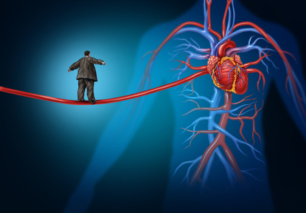
Blood Supply
The blood supply to the brain comes from the internal carotid and vertebral arteries, lying in the subarachnoid space. The internal carotid arteries branch from the common carotid arteries (that can be felt pulsing in the neck) and enter the head. Each internal carotid artery ascends to the level of the optic nerve, where each divides into three branches: the ophthalmic artery, anterior cerebral artery that supplies the frontal and parietal lobes and a middle cerebral artery that supplies the midbrain and lateral surfaces of the cerebral hemispheres. The vertebral arteries start at the base of the neck and pass upward through holes in the side of the vertebral bones of the neck. After ascending to the brain, the two vertebral arteries fuse to form the basilar artery.
The basilar artery goes up the front surface of the pons, sending off many branches, before dividing into the posterior cerebral arteries. The vertebral arteries and their branches supply the brain posterior to the area supplied by the internal carotids. To keep flexibility in the circulatory system, there are many connections between the different blood supplies, and these connections form a loop that is known as the circle of Willis. The circle is made whole by smaller arteries such as the posterior communicating artery and anterior communicating artery. The purpose of this is that if one of the arteries becomes blocked, or blood supply is cut off for whatever reason, then blood supply can be increased from one of the other arteries to compensate.
Organisation of the Brain
As can be gathered from the above information, the brain is a complicated organ! However, the structure alone does not even begin to scratch the surface of the complexity of the brain. Within each lobe and each section, there are many more functional areas. In some areas however, there are interesting patterns that serve to illustrate the underlying order of the brain as a whole. As has been discussed before, the motor area lies within the frontal lobe, in a gyrus called the pre-central gyrus. Within this gyrus, there are actually parts of the gyrus that each correspond to a certain section of the body. The way this is organised is actually as if a man is lying over the gyrus, with his hands outstretched above his head. Imagine this man lying along the length of the gyrus (and his twin on the other side), sprawling away from the centre. This corresponds to the areas that actually control those areas of the brain, so that the areas that control the hands lie next to those of the arms, which lie just before we reach the head, passing down the body until we reach the feet at the other end of the gyrus. However, as some areas of the body require lots of information to be sent to them regarding motor control (especially the hands, for example), they take up far more of the brain than they do in actual body terms. The same applies to the sensory part of the brain.
Other parts of the brain are also well ordered, vision for example is actually broken down into different elements of what it means to see. Movement can be dealt with separately from shapes, which can be dealt with separately to depth perception etc. And then there is a separate area that actually deals with interpreting these shapes and deciding what the most likely object is. Due to all this complexity, the brain is still something of a mystery. We still do not fully understand many functions of the brain, such as how we are able to exert free-will, how memories are stored and what causes our personalities; however, perhaps it is these unknowns that keep the brain as such a fascinating object.
More information
 |
For more information on brain health, including nutrition, exercise and mental activity, see Brain Health. |
References
- Graybiel AM. The Basal Ganglia: learning new tricks and loving it. Current Opinion in Neurobiology. 2005;15:638-44.
- Martini FH, Timmons MJ, McKinley MP. Human Anatomy. Third ed. Upper Saddle Creek: Prentice Hall 2000.
- Moore KL, Dalley AF. Clinically Oriented Anatomy. Philidelphia: Lippincott Williams & Wilkins 1999.
- Nolte J. The Human Brain. Fifth ed. St Louis: Mosby 2002.
- Pritchard TC, Alloway KD. Medical Neuroscience. First ed. Madison: Fence Creek Publishing 1999.
All content and media on the HealthEngine Blog is created and published online for informational purposes only. It is not intended to be a substitute for professional medical advice and should not be relied on as health or personal advice. Always seek the guidance of your doctor or other qualified health professional with any questions you may have regarding your health or a medical condition. Never disregard the advice of a medical professional, or delay in seeking it because of something you have read on this Website. If you think you may have a medical emergency, call your doctor, go to the nearest hospital emergency department, or call the emergency services immediately.
Related Articles


Need a health appointment?
Find and book a doctor, dentist, physio and more on Healthengine
Find a practitioner