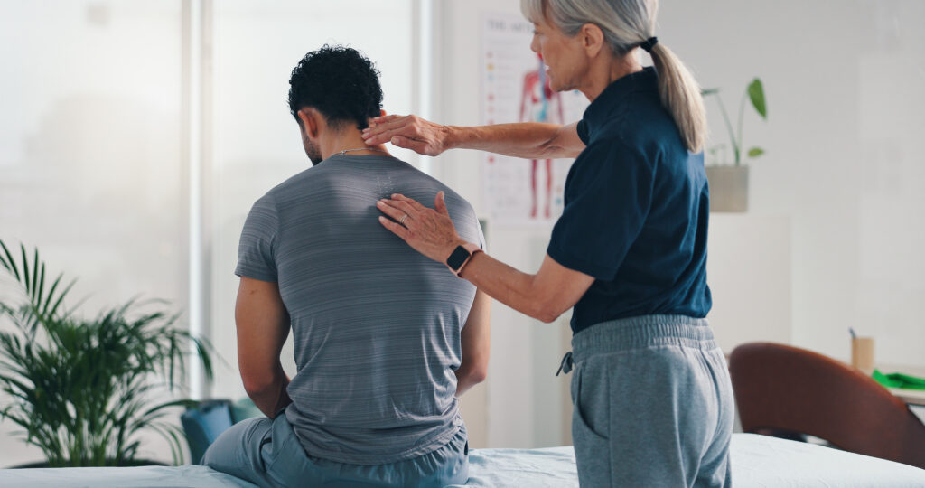
- An introduction to the anatomy of the shoulder
- Bony anatomy of the shoulder
- Shoulder ligaments, labrum, bursae and the joint capsule
- Muscles and tendons of the shoulder
- Injury to the shoulder
An introduction to the anatomy of the shoulder
| The shoulder girdle is mainly made up of the true shoulder joint (glenohumeral joint) and the joint between the shoulder blade and the chest (scapulothoracic joint). When we move our arm around, movement occurs at both of these joints, with most (two thirds) occurring at the shoulder joint. The shoulder is basically like a ball and socket. The “ball” is the head of the humerus and the “socket” is theglenoid part of the shoulder blade (scapula).The anatomy of the shoulder is unique – it has a relatively shallow socket which results in amazing flexibility and range of motion to the shoulder joint which is unparalleled elsewhere in the body. In order to achieve this flexibility but maintain a stable shoulder, there is a complex interplay between the joints, muscles and ligaments. Injury to any one of these structures can therefore result in significant ongoing pain, weakness, or instability. |
Bony anatomy of the shoulder
The humeral head is the “ball” part of the ball and socket. It is made up 4 of components:
- Rounded joint surface – the head of the humerus which sits in the socket (glenoid) to form the shoulder joint.
- A tuberosity is a distinct prominence or bump on a bone which is usually where a muscle or a ligament attaches. There are two of these on the humerus, the greater tuberosity and the lesser tuberosity. The greater tuberosity is the site of insertion for three important shoulder (rotator cuff) muscles; the supraspinatus, infraspinatus and teres minor. The lesser tuberosity is the site of subscapularis insertion which is another shoulder muscle.
- The bicipital groove is the groove between the greater and lesser tubercles. The groove functions as a passage for the tendon of the long head of the biceps brachii.
The scapula (shoulder blade)

The scapula (commonly known as the shoulder blade) is a triangular shaped bone which has a part called the glenoid that forms the “socket” for the head of the humerus. The glenoid cavity is not even half the size of the humeral head so it does not provide the joint with much stability. However, the glenoid socket is made deeper by a rim of “gristle” (fibrocartilage) called the labrum that runs around the periphery of the socket, effectively doubling its depth. When somebody dislocates their shoulder for the first time, they usually rip off this gristle, which then makes their socket shallower and predisposes them to repeat shoulder dislocations. The ball and socket of the shoulder are enclosed by a “sock” of tissue, known as the joint capsule. If this sock of tissue becomes stiff, it can result in shoulder stiffness and pain known as frozen shoulder (adhesive capsulitis).
The shoulder blades sit on the upper part of the back (thorax), with one on each side. Projecting from the front of the shoulder blade is a hook of bone known as the coracoid process. The coracoid process is a bone which serves as the connection point for the ligaments that connect the shoulder blade to the collarbone. Another large hook or shelf of bone projects upwards from the shoulder blade and is known as the acromion. The acromion serves as an attachment for the strong and important deltoid muscle.
The clavicle (commonly known as the collar bone) connects the shoulder to the rest of the body via two joints, the acromioclavicular joint and the sternoclavicular joint.
The acromioclavicular joint (AC joint) is at the tip of the shoulder and is the site where the collarbone connects to the shoulder blade. The collarbone serves to strut the shoulder blade out from the body, allowing the surrounding muscles to work at their most effective tensions and generate more power. The AC joint commonly wears out as we get older resulting in arthritis, which may or may not be painful.
The sternoclavicular joint is at the base of the neck and is a saddle joint which sits between the collar bone and the breast plate (sternum). This joint also helps to strut the shoulder blade out from the body.
Book your health appointments online
Find and instantly book your next health appointment with Healthengine
Shoulder ligaments, labrum, bursae and the joint capsule
The ligaments, joint capsules and labrum are fixed structures that stabilise and reinforce the shoulder.
The glenoid labrum is a rim of gristle (fibrocartilage) attached to the periphery of the glenoid cavity and acts to deepen the socket and increase the contact surface area between the ball and socket, increasing the stability of the shoulder joint.
The shoulder requires a large number of bursae. Bursae are fluid filled sacs that roll over themselves and serve to lubricate the points of possible friction between the muscles and the joint capsule. The major bursae in the shoulder include the subacromial bursa, the subdeltoid bursa and the subscapular bursa.The subacromial bursa can commonly become inflamed and cause pain (bursitis). It is a common site for injections of cortisone to help decrease the pain in the shoulder.
There are three glenohumeral ligaments which provide some support to the front of the shoulder joint; the superior, middle and inferior glenohumeral ligaments. The superior glenohumeral ligament works in conjunction with the coracohumeral ligament to stabilise the humeral head. The middle glenohumeral ligament is not always present. When it is present, it aids to stabilise the humeral head. The thick inferior ligament is the primary stabiliser of the three.
Muscles and tendons of the shoulder
The muscles are the “fleshy” components of the body that contract to effect movement by pulling on tendons, that are the “sinew” that connects the muscles to the bones.
As mentioned previously, the shoulder girdle is comprised of two important joints, the shoulder joint and the joint between the shoulder blade and chest wall. Each of these joints has muscles which help to move them.
Shoulder joint muscles (glenohumeral joint)
The shoulder joint has very large powerful muscles which provide the power for strong movements (deltoid, pec major), and smaller muscles (rotator cuff) which help with stability of the glenohumeral joint. If a very powerful muscle like the deltoid contracts on its own, the humeral head would partially slip out of place (sublux) due to the shallowness of the socket. This is prevented by the rotator cuff muscles which co-contract with the deltoid, compressing the humeral head into the glenoid and providing the dynamic stability required for strong shoulder movements.The muscles which comprise the rotator cuff and their individual motions are:
- Supraspinatus – assists with lifting the arm above the head. This is the most common muscle / tendon to tear in the shoulder.
- Subscapularis – twists the arm behind the back.
- Infraspinatus and the teres minor – twist the arm out sideways from the body.
It is important to understand that the rotator cuff functions together as a unit to compress the ball into the socket.
- The deltoid muscle is the “powerhouse” and the most important muscle of the shoulder. The normal functioning of this muscle is critical for overhead shoulder activity.
- The pectoralis major muscle is another powerful muscle which like the deltoid is integral to shoulder function. We use this muscle when doing push-ups. It originates from the front of the chest and collar bone and inserts on the upper part of the arm bone (humerus).
- The latissimus dorsi is another powerful muscle that together with the teres major muscle, pulls the arm down to the side. We use this muscle when doing chin-ups.
Muscles that move the shoulder blade on the chest wall (scapulothoracic articulation)
- The trapezius forms the “webbing” of the neck, and runs from the spinal column out to the shoulder blade and collar bone. It helps to shrug the shoulders.
- The rhomboids and levator scapulae are small muscles that join the shoulder blade to the spinal column.
- The serratus anterior muscle helps to stabilise the shoulder blade on the chest wall. When it is not functioning, “winging” occurs which is when the shoulder blade protrudes from the back. This muscle inserts into the shoulder blade and originates from the first 9 ribs.
Injury to the shoulder
Shoulder injuries are common accounting for up to 20% of all athletic injuries. As mentioned previously, the unique structure of the shoulder joints results in a multiaxial universal joint with an unparalleled range of motion. However, this unique structure also means that the shoulder joint is the most frequently dislocated joint in the body. In addition to shoulder dislocations, other common injuries include rotator cuff tendon tears and broken bones including the humerus and collar bone.
Kindly reviewed by:
Dr John Trantalis
MBBS FRACS
Specialist Surgeon of Shoulder & Elbow Surgery at the Orthopaedic and Joint Replacement Centre, NSW; and Editorial Advisory Board Member of the Virtual Bone and Rheumatology Centres.
References
- Marieb EN, Hoehn K. Chapter 8: Joints. Anatomy and Physiology. 3rd ed. United States: Pearson Benjamin Cummings; 2007: 239-40.
- Terry GC, Chopp TM. Functional Anatomy of the Shoulder. J Athlet Train. 2000; 35(3): 248-55.
- Martini FH. Chapter 9: Articulations. Fundamentals of Anatomy and Physiology 4th ed. New Jersey, Prentice Hall International; 1998: 267-8.
All content and media on the HealthEngine Blog is created and published online for informational purposes only. It is not intended to be a substitute for professional medical advice and should not be relied on as health or personal advice. Always seek the guidance of your doctor or other qualified health professional with any questions you may have regarding your health or a medical condition. Never disregard the advice of a medical professional, or delay in seeking it because of something you have read on this Website. If you think you may have a medical emergency, call your doctor, go to the nearest hospital emergency department, or call the emergency services immediately.
Related Articles
The reality of living with diabetes, and how…

Endometriosis is a condition that approximately 700,000 plus…

While it may be easy to dismiss children’s…
Need a health appointment?
Find and book a doctor, dentist, physio and more on Healthengine
Find a practitioner