
Carotid Doppler Ultrasound Scan
What is a carotid doppler ultrasound?
A carotid doppler ultrasound is primarily for assessing the blood flow in the arteries of the neck.
How is a carotid doppler ultrasound performed?
A carotid doppler is a special type of ultrasound which is able to show blood flow as a colour-coded dynamic image. The sonographer is able to calculate the velocity of blood flow and to determine the degree of narrowing in the blood vessel, if any.
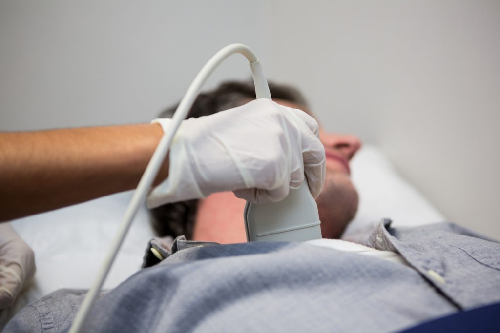
The test consists of applying a small probe to the side of the neck, near the Adam’s apple, using sonographic gel applied to the skin. Mild to moderate pressure may be necessary to obtain detailed images of the carotid arteries.
Why would you need a carotid doppler ultrasound?
A carotid doppler ultrasound is usually performed to assess the risk of stroke in a patient with episodes suggestive of cerebro-vascular disease, such as a transient ischaemic attack (TIA) – also known as a mini-stroke.
A TIA is caused by a small clot temporarily blocking off an area of blood supply in the brain. This may result in a range of sensory symptoms, such as numbness, tingling, or altered sensation, or motor symptoms – weakness of the face, arm or leg, commonly on one side of the body. A clot in any blood vessel supplying the posterior part of the brain, the cerebellum, may cause unsteadiness, clumsiness, slurred speech or even loss of consciuosness. This is known as a posterior-circulation TIA.
If the clot is dissolved by the body’s natural processes within a few minutes, blood flow returns to the affected area of the brain, and function returns to normal. Otherwise, there may be varying degrees of residual symptoms, with variable rates of recovery, right up to a completed infarction – a ‘full-blown’ stroke.
The carotid arteries in the neck are a common source of small clots, especially if there is a narrowing of the blood vessel due to atherosclerosis, known as a carotid artery stenosis. This narrowing may be detected by your doctor as a carotid bruit – a ‘whooshing’ noise due to turbulent flow, heard with a stethoscope placed on the side of the neck, over the carotid artery.
Test results explained
The carotid doppler ultrasound results may be reported as normal, meaning normal blood flow and no evidence of carotid stenosis (narrowing).
When there is some narrowing of the vessel, this usually reported in terms of a percentage, for example 70% stenosis.
With stenosis over 75%, your doctor may refer you to a vascular surgeon for consideration of surgery to correct the narrowing, known as a carotid end arterectomy (CEA). Lesser degrees of stenosis are usually treated with a daily low dose of aspirin to decrease the likelihood of clot formation.
Related specialists
Related tests
Also known as
- Carotid US
- Carotid U/S
- Carotid USS
- Carotid Duplex Doppler
Links
A: Use HealthEngine to find and book your next Vascular Surgeon appointment. Click on the following locations to find a Vascular Surgeon clinic in your state or territory.
This article is for informational purposes only and should not be taken as medical advice. If in doubt, HealthEngine recommends consulting with a registered health practitioner.
All content and media on the HealthEngine Blog is created and published online for informational purposes only. It is not intended to be a substitute for professional medical advice and should not be relied on as health or personal advice. Always seek the guidance of your doctor or other qualified health professional with any questions you may have regarding your health or a medical condition. Never disregard the advice of a medical professional, or delay in seeking it because of something you have read on this Website. If you think you may have a medical emergency, call your doctor, go to the nearest hospital emergency department, or call the emergency services immediately.
Related Articles


Need a health appointment?
Find and book a doctor, dentist, physio and more on Healthengine
Find a practitioner